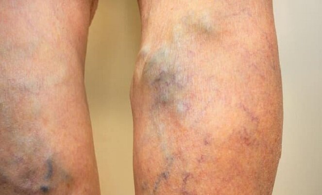
According to reports ImmenaResearchers at Pennsylvania State University have announced in a recent study a new imaging technique for identifying and treating blood clots. Thrombosis Deep vein (DVT) means the formation of a blood clot in the inner wall of a deep vein. In some cases, failure to treat this is dangerous and can cause Embolism The lungs become fatal.
for management Thrombosis Deep vein occlusion and rapid detection, monitoring and treatment are critical. Current diagnostic imaging techniques do not have the clarity to pinpoint the exact location of a blood clot and the ability to monitor clots instantly. Ultrasound is also commonly used for diagnosis Thrombosis A vein is used, but for diagnosis Thrombosis Deep vein is not a good method.
This method can show doctors the existence of a strange fluid flow, but it is not possible to determine exactly what it is. Now it may be related to a blood clot and it may be related to something else. Therefore, patients should also have a blood test to find out.
The doctor may prescribe medication after diagnosing those factors and eliminate them, or he may decide to use a tool to go inside the body and catch and remove the clot. In addition, the drugs have disadvantages because they may not be able to eliminate the clot or cause bleeding in other parts of the body.
To better identify the location of blood clots, researchers at Pennsylvania State University have announced a new technology that can locate and treat blood clots. The researchers in this study used a method called “Nanoptysis(NPeps) used.
In this method, the particles are based on a crust around a drop of oil Florin Which are similar to liquid Teflon. The surface of the shell contains a molecule to which it attaches as soon as the protein on the surface of the activated platelets (except the main cell of the blood clot) is identified.
“squat Medina“These particles attach to the surface of the clots, and after we apply the ultrasound, they turn to gas, creating a bubble under the shell, which is a great contrast for imaging,” said Scott Medina, a professor of medical engineering. These bubbles appear exactly where the clots form.
The researchers injected the enzyme that causes blood clots into cows to analyze how to diagnose and treat clots. The enzyme caused blood clots to form 100 percent of the time, but that happened 30 percent of the time when those particles came into action. We were surprised because it was as if these particles were not only attaching to the clots, but also somehow breaking them down.
These particles are thought to saturate the surface of the blood clot and inhibit further blood clot growth mechanisms, and after ultrasound, the particles disrupt the function of the blood clot and continue to inhibit its mechanism.
Researchers will do more research in the future to determine how these particles disrupt clots and how they can have more control over the behavior of these particles.
Source: ISNA
Comments
Post a Comment